Tendon Diagram : 1. Cyst on the lower part of the diagram. Tendons are found throughout the body, from the head and neck all the way down to the feet. Ligaments and tendons are fibrous connective tissues made up of densely packed collagen fibers. Ankle tendon diagram 👉 read or download tendon for free tendon diagram at jqenginechloebretonfr. The bones together make up the hip.
ads/bitcoin1.txt
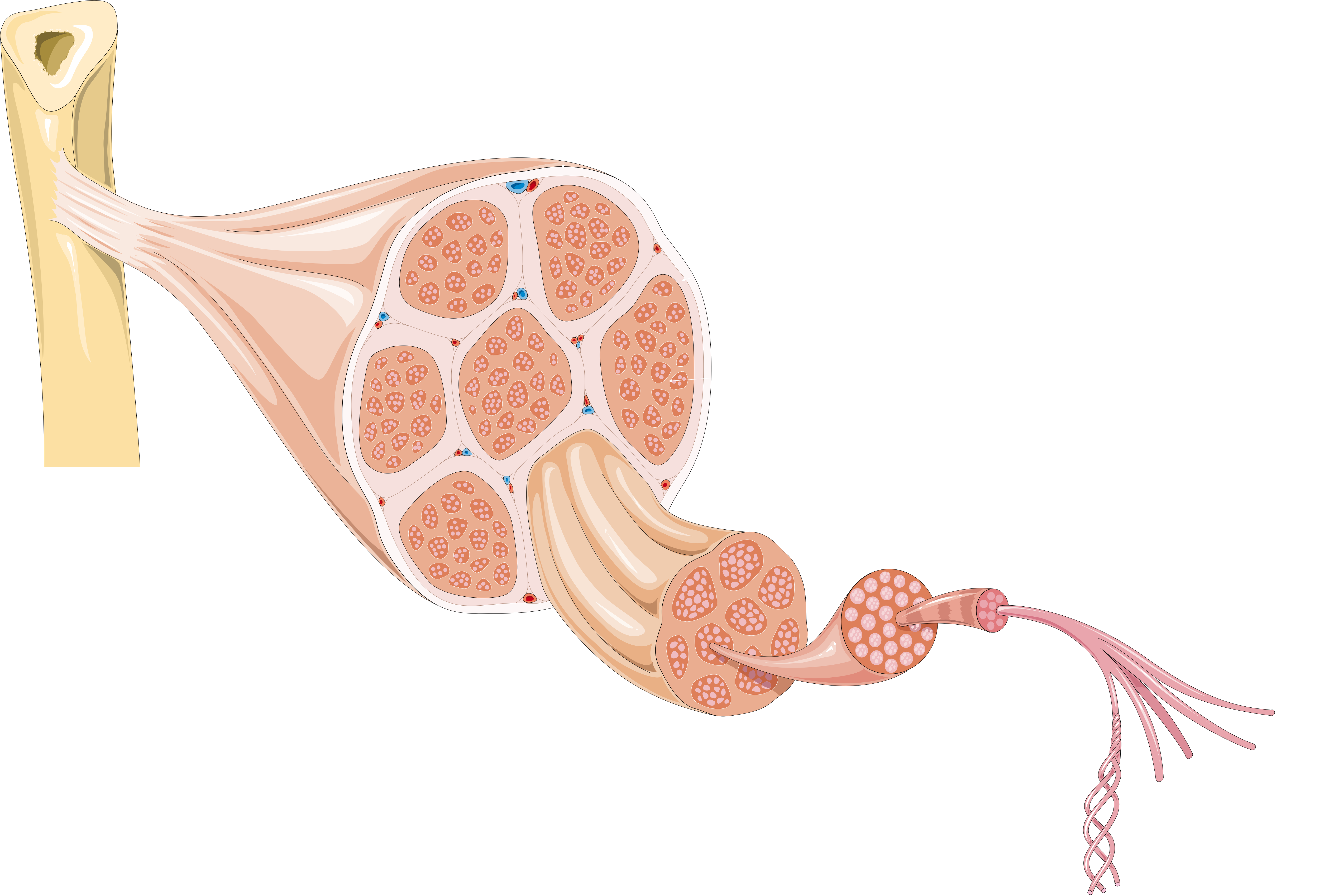
Tendons are thick bands of tissue that connect muscles to bones. You can see a diagram of the achilles tendon below. Learn about the anatomy and physiology of tendons. The pubis, ischium, and ilium together constitute the pelvis while the thigh bone is the femur. The muscles that make up the quadriceps are the strongest and leanest of all muscles in the body.
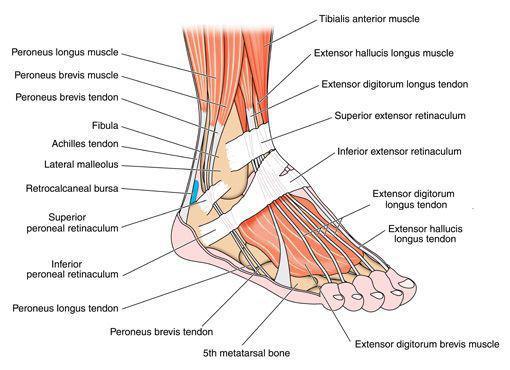
Knee Tendons Medical Vector Illustration Scheme Anatomical Diagram Stock Illustration Download Image Now Istock from media.istockphoto.com A change in shape of a muscle (the stimulus) causes the muscle to readjust its shape (the response) and maintain your posture. Tendons are found throughout the body, from the head and neck all the way down to the feet. The anterior tibial tendon allows us to raise the foot. By connecting our rigid bones to our powerful muscles, tendons allow us to move. The two peroneal tendons in the foot run side by side behind the outer ankle bone. Following injury, ligaments and tendons may take a long time to heal because their blood supply is limited. Learn vocabulary, terms and more with flashcards, games and other study tools. Ligaments join the knee bones and provide stability to the knee:
Diagram of a catheter in the neck.
ads/bitcoin2.txt
The bones of the hip include the femur, the ilium, the ischium, and the pubis. Feet human anatomy bones tendons ligaments and more. Cyst on the lower part of the diagram. Tendons are found throughout the body, from the head and neck all the way down to the feet. The bones together make up the hip. Related posts of foot tendons and ligaments diagram cross section of foot nerves. Allows the foot to be turned inward and also supports the arch of the foot. One peroneal tendon attaches to the outer part of the midfoot, while the other tendon runs under the foot and attaches near the inside of the arch. By connecting our rigid bones to our powerful muscles, tendons allow us to move. Allows the action of raising the foot. The long head of biceps (lhb) is a very important tendon that travels through the shoulder joint (glenohumeral joint).the biceps tendon begins at the top of the shoulder socket (the glenoid) and then passes across the front of the shoulder to connect to the biceps muscle. Ligaments and tendons are fibrous connective tissues made up of densely packed collagen fibers. 1 article features images from this case.
Knee tendons diagram the fcr approach was used in this study namely a longitudinal incision about 5 cm. Below is a diagram of the hamstring tendon. Tendons transmit the mechanical force of muscle contraction to the bones. One peroneal tendon attaches to the outer part of the midfoot, while the other tendon runs under the foot and attaches near the inside of the arch. A body muscle diagram is used by different people for various uses.
File Tendon Anatomy 1 Smart Servier Png Wikimedia Commons from upload.wikimedia.org The achilles tendon is also called the calcaneal tendon. The muscles that make up the quadriceps are the strongest and leanest of all muscles in the body. Learn about the anatomy and physiology of tendons. Tendons are found throughout the body, from the head and neck all the way down to the feet. Diagram of the ankle bones. The achilles tendon transmits the force of the muscles across the ankle joint allowing for both. The achilles tendon enables us to walk, without it we would not be able to raise our heels of the ground. Check out and click on the image to download it.
A body muscle diagram is used by different people for various uses.
ads/bitcoin2.txt
The pubis, ischium, and ilium together constitute the pelvis while the thigh bone is the femur. Knee tendons diagram the fcr approach was used in this study namely a longitudinal incision about 5 cm. The long head of biceps (lhb) is a very important tendon that travels through the shoulder joint (glenohumeral joint).the biceps tendon begins at the top of the shoulder socket (the glenoid) and then passes across the front of the shoulder to connect to the biceps muscle. The anterior tibial tendon allows us to raise the foot. The diagram below shows how this reflex works. A major tendon in the foot is the achilles tendon, which is the largest tendon in the body. This important tendon in the back of the calf and ankle connects the plantaris, gastrocnemius, and soleus muscles to. A tendon is a band of tissue that connects a muscle to a bone. The bones together make up the hip. To bend the elbow and to turn the palm of the hand towards the sky. Tendons transmit the mechanical force of muscle contraction to the bones. For example, a tap to the tendon under the knee cap elicits (triggers) the knee jerk reflex. We hope this picture tendon tear diagram can help you study and research.
You can see a diagram of the achilles tendon below. The two peroneal tendons in the foot run side by side behind the outer ankle bone. The achilles tendon is the strongest and largest tendon in the body. Muscles in your body diagram. Movement occurs when our muscles pull on our bones, relocating them.
Foot And Ankle Sportsmed from sportsmedalabama.com Posted on april 3, 2019april 3, 2019. Diagram of the ankle bones. The achilles tendon is the strongest and largest tendon in the body. Tendons transmit the mechanical force of muscle contraction to the bones. By connecting our rigid bones to our powerful muscles, tendons allow us to move. Biceps tendons the biceps muscle has two tendons at the shoulder, called the long head and short head. Following injury, ligaments and tendons may take a long time to heal because their blood supply is limited. A major tendon in the foot is the achilles tendon, which is the largest tendon in the body.
Tendon, tissue that attaches a muscle to other body parts, usually bones.
ads/bitcoin2.txt
Also allows the action of raising up onto toes. Posted on april 3, 2019april 3, 2019. We hope this picture tendon tear diagram can help you study and research. Ligaments join the knee bones and provide stability to the knee: The two peroneal tendons in the foot run side by side behind the outer ankle bone. A body muscle diagram is used by different people for various uses. The changes in ligaments and tendons generally occur more slowly than adaptation in bone, because ligaments and tendons have less vascular supply. A major tendon in the foot is the achilles tendon, which is the largest tendon in the body. Diagram of inside the body. Muscles in your body diagram. The bones of the hip include the femur, the ilium, the ischium, and the pubis. Allows the foot to be turned inward and also supports the arch of the foot. Movement occurs when our muscles pull on our bones, relocating them.